Clinical Features of Myasthenia Gravis:
 Myasthenia gravis is a disorder of neuromuscular transmission characterised by:
- Weakness and fatiguing of some or all muscle groups
- Weakness worsening on sustained or repeated exertion, or towards the end of the day, relieved by rest
This condition is a consequence of an autoimmune destruction of the NICOTIN1C POSTSYNAPTIC RECEPTORS FOR ACETYLCHOLINE.
Myasthenia gravis is rare, with a prevalence of 5 per 100 000. The increased incidence of autoimmune disorders in patients and first degree relatives and the association of the disease with certain histocompatibility antigens (HLA) – B7, B8 and DR2 – suggests an IMMUNOLOGICAL BASIS.
Antibodies bind to the receptor sites resulting in their destruction (complement mediated). These antibodies are referred to as ACETYLCHOLINE RECEPTOR ANT1BODIES (AChR antibodies) and are demonstrated by radioimmunoassay in the serum of 90% of patients.
Muscle biopsy may show abnormalities:
- Lymphocytic infiltration associated with small necrotic foci of muscle fibre damage.
- Muscle fibre atrophy (type I and II or type III alone).
- Diffuse muscle necrosis with inflammatory infiltration (when associated with thymoma)
Motor point biopsy may show abnormal motor endplates. Supravital methylene blue staining reveals abnormally long and irregular terminal nerve branching. Light and electron microscopy show destruction of ACh receptors with simplification of the secondary folds of the postsynaptic surface.
Clinical Features
Up to 90% of patients present in early adult life (<40 years of age). Female: male ratio 2:1. The disorder may be selective, involving specific groups of muscles.
Several clinical subdivisions are recognised:
- Class 1 – ocular muscles only – 20%
- Class 2 – Mild generalised weakness
- Class 3 – Moderate generalised and mild to moderate ocular-bulbar weakness
- Class 4 – Severe generalised and ocular-bulbar weakness
- Class 5 – Myasthenic crises
Approximately 40% of class I will eventually become widespread. The rest remain purely ocular throughout the illness. Respiratory muscle involvement accompanies severe illness.
Clinical Features
Cranial nerve signs and symptoms
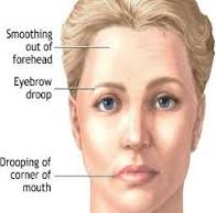
- Ocular involvement produces ptosis and muscle paresis.
- Weakness of jaw muscles allows the mouth to hang open.
- Weakness of facial muscles results in expressionless appearance.
Braner Pain Clinics has a talented and friendly staff. We will do everything in our power to make sure your visit is a satisfying experience. If there is anything else you may need from us, just ask! We are here to serve you.