How do you get relief from Parkinson’s Disease?

Clinical Features , Diagnosis And Treatment of Parkinson’s Disease:Parkinson’s Disease
AETIOLOGY                                 

The causes of Parkinson disease are unknown. Gene mutations have been identified in young onset and familial cases (synuclein, parkin and LRRK2).

Parkinson disease in humans and animals has resulted in increased interest in the role of toxins and an animal model for developing new treatments.

Parkinsonian features may be present in many disorders and are not always treatment (L Dopa) responsive. These disorders usually share features of slowness and rigidity (kinetic rigid syndromes)

1.Parkinson’s disease                    

  • Mimics  – Multiple system atrophy (MSA)
  • Progressive supranuclear palsy (PSP)
  • Corticobasal ganglionic degeneration
  • Diffuse Lewy body disease (DLBD)

2. Secondary Parkinsonism

  • Drug induced (dopamine receptor blockers-antipsychotics/antiemetics; sodium valproate)
  • Post traumatic (pugilists encephalopathy)
  • Vascular disease (small vessel multi-infarct state)
  • Infectious (post encephalitic/prion disease/HIV)
  • Miscellaneous: hydrocephalus/parathyroid/paraneoplastic

Pathology of Idiopathic

The substantia nigra contains pigmented cells (neuromelanin) which give it a characteristic ‘black’ appearance (macroscopic). These cells are lost in Parkinson’s disease and substantia nigra becomes pale. Remaining cells contain atypical eosinophillic inclusions in the cytoplasm. Lewy bodies may be found in the cerebral cortex especially when dementia is present (diffuse Lewy body disease). Changes are seen in other basal nuclei striatum and globus pallidus.

CLINICAL FEATURES

  • Initial symptoms are vague, the patient complains of aches and pains.
  • A coarse TREMOR at a rate of 4-7 Hz usually develops early in the disease.
  • It begins unilaterally in the upper limbs and eventually spreads to all four limbs.
  • The tremor is often ‘pill rolling’, the thumb moving rhythmically backwards and forwards on the palm of the hand.
  • It occurs at rest, improves with movement and disappears during sleep.
  • RIGIDITY is detected by examination. It predominates in the flexor muscles of the neck, trunk and limbs and results in the typical ‘flexed posture’.

BRADYKINESIA: This slowness or paucity of movement affects facial muscles of expression (mask-like appearance) as well as muscles of mastication, speech, voluntary swallowing and muscles of the trunk and limbs. Dysarthria, dysphagia and a slow deliberate gait with little associated movement (e.g. arm swinging) result.

  • Tremor, rigidity and bradykinesia deteriorate simultaneously, affecting every aspect of the patient’s life:
  • Handwriting reduces in size.
  • The gait becomes shuffling and festinant (small rapid steps to ‘keep up with’ the centre of gravity) and the posture more flexed.
  • Rising from a chair becomes laborious with progressive difficulty in initiating lower limb movement from a stationary position.
  • Eye movements may be affected with loss of ocular convergence and upward gaze.
  • Excessive sweating and greasy skin (seborrhea) can be troublesome.
  • Depression occurs in about 50%.
  • As the disease progresses the frequency of drug induced confusional states and dementia increases, with 80% developing dementia after 20 years of disease (if they survive).
  • Autonomic features occur – postural hypotension, constipation.
  • REM sleep behavior disorder – where patient acts out dreams and may hurt themselves or their sleep partner. May precede onset of motor symptoms.
  • Time of onset is mid-late fifties with increasing incidence with increasing age. Juvenile presentation can occur, when presentation and disease progression is often atypical; a genetic basis is more often found.

DIAGNOSIS

The diagnosis of PD in the early stages is difficult. Post-mortem data from the London Brain Bank shows this to be incorrect in 25% of those diagnosed in life.

  • New tremor in middle age causes particular difficulty – senile/essential & metabolic tremor generally absent at rest and worsened by voluntary movement.
  • The diagnostic use of a L-dopa or dopamine agonist (apomorphine) challenge has decline due to concerns that it may increase the risk of subsequent drug induced dyskinesia.
  • Functional imaging (SPECT & PET) should improve diagnostic accuracy and ensure that persons with conditions unresponsive to treatments (PD mimics) are not unnecessarily exposed to them.

TREATMENT

Treatment is symptomatic and does not halt the pathological process. No agents have yet demonstrated convincing neuroprotective effect.

  • Levo dopa is given with a decarboxylase inhibitor, which prevents peripheral breakdown in the liver (as in I) allowing a higher concentration of dopa to reach the blood-brain barrier (as in 2) and reduces the peripheral side effects (nausea, vomiting, hypotension).
  • Central side effects: confusion, depression, dyskinetic movements and following long-term treatment – ‘On/Off’ phenomenon.
  • Rapid onset or longer action can be achieved using dispersible or controlled-release preparations.
  • Exogenous dopa improves bradykinesia, rigidity and, to a lesser extent, tremor, but in 20% the response is poor. Dopa has relatively less effect on non-motor symptoms.
  • A new preparation of dopa is available for continuous infusion via jejunostomy in severe disease.
  • Dopamine agonists: Now used earlier in disease management, they act directly on the dopamine receptor independent of degenerating dopaminergic neurons. It is not clear if patients do better in the long term if dopamine agonists or dopa are used first. There are two types of dopamine agonists, ergot derived, including pergolide, cabergoline, apomorphine and non-ergot derived.
  • COMT inhibitors: Entacapone reduces the metabolism of levodopa and is used as adjunctive treatment. Tolcapone is an alternative that can cause hepatic toxicity; it requires close monitoring.

For you, the patient or carer, we hope our website www.branerpainclinic.com covers any question you may have regarding Parkinson’s disease in your life. However, if you can’t find quite what you’re looking for – or you would like to know more – please feel free to Contact Us at our clinic Braner Pain Clinic and Call Now : 1 (877) 573-1282. We’d be glad to help in any way we can.
How to treat Vascular Malformations in the Brain?

Investigations, Management And Treatment of Vascular Malformations:Vascular Malformations

Vascular malformations vary in size and different forms exist:

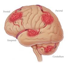
Arteriovenous malformations (AVMs) are developmental anomalies of the intracranial vasculature; they are not neoplastic despite their tendency to expand with time and the descriptive term ‘angioma‘ occasionally applied.

Dilated arteries feed directly into a tangled mass of blood vessels of varying calibre; they bypass capillaries and shunt oxygenated blood directly into the venous system. Due to high intraluminal pressure, veins may adopt an ‘aneurysmal’ appearance. Arteriovenous malformations occur at any site but are commonest in the middle cerebral artery territory.

  • Capillary Telangiectasis:  An area of dilated capillaries, like a small petechial patch on the brain surface – especially in the pons. These lesions are often only revealed at autopsy.
  • Cavernous Malformation/Angioma: Plum colored sponge-like mass composed of a collection of blood filled spaces with no intervening brain tissue. No enlargement of feeding or draining vessels.

ARTERIOVENOUS MALFORMATIONS

CLINICAL PRESENTATION

  • Haemorrhage: About 40 60% of patients with an AVM present with haemorrhage -often with an intracerebral or intraventricular component. In comparison with saccular aneurysms, AVMs tend to bleed in younger patients, i.e. 20-40 years, and are less likely to have a fatal outcome. Vasospasm and delayed ischaemic complications rarely develop. Small AVMs, those with high intranidal pressure and those draining exclusively to deep veins have an increased risk of haemorrhage.

1)      Annual Risk of Haemorrhage: Patients with no history of haemorrhage have an annual risk of bleeding of 2-4%. For those presenting with haemorrhage, the risk of re-bleeding may be higher, particularly in the first year. One study reported an annual risk of 17%.

2)      Mortality from Haemorrhage: In contrast to the high mortality following aneurysm rupture, haemorrhage from an AVM carries the relatively low mortality rate of approximately 10%.

  • Epilepsy: Generalized or partial seizures commonly occur in patients with arteriovenous malformation, especially if the lesion involves the cortical surface. Of patients presenting with haemorrhage, 30% have a history of epilepsy.
  • Neurological Deficit: Large AVMs, especially those involving the basal ganglia, may Present with a slowly progressive dementia, Hemiparesis or visual field defect, Probably as a result of a ‘steal’ effect. The infrequent brain stem AVM may also produce a motor or sensory deficit, with or without cranial nerve involvement.
  • Headache: Attacks of well localized headache – unilateral and throbbing – occur in a Proportion of patients subsequently shown to have a large AVM.
  • Cranial Bruit: Auscultation, especially over the eyeball, occasionally reveals a bruit.

INVESTIGATIONS

  • CT scan: Most AVMs are evident on CT scan unless masked by the presence of an intracranial haematoma. A double dose of intravenous contrast may aid visualization, especially with small cryptic lesions.
  • MRI: Conventional MRI will clearly demonstrate the AVM as a region of flow voids, with associated signal change within or around the lesion from areas of old haemorrhage or gliosis. The MRI provides exact anatomical detail and helps surgical Planning. Functional MRI aids identification of any adjacent eloquent areas.
  • Angiography: Both CT and MR angiography should confirm the presence of an AVM but digital subtraction four-vessel angiography is required to delineate the feeding and draining vessels. Occasionally small AVMs are difficult to detect and only early venous filling may draw attention to their presence.
  • N.B.: In the presence of a haematoma, digital subtraction angiography should be delayed until the haematoma resolves, otherwise local pressure may mask demonstration of an AVM. If the angiogram is subsequently negative, then MRI is required to exclude the presence of cavernous malformation.

MANAGEMENT

Various methods of treating arteriovenous malformations are available. All risk further damage and a team comprised of the neurosurgeon and neuroradiologist should decide on the optimal method or combination of methods for each patient. The urgency of the patient’s clinical condition and the risks of treatment must be weighed against the risk of a conservative approach.

Indications for Intervention

  • Expanding haematoma associated with AVM
  • Progressive neurological deficit
  • Risk of haemorrhage especially
  • young patients with many years at risk
  • AVMs < 3 cm

METHODS OF TREATMENT

  • Operation:  Excision – complete excision of the AVM (confirmed by per- or postoperative angiography) is the most effective method of treatment particularly for small AVMs in non-eloquent areas. Image guidance may aid localization. Larger lesions (> 6 cm) have a greater risk of postoperative hyper perfusion syndrome and brain swelling and carry a 40% risk of permanent neurological deficit.
  • Stereotactic Radio-Surgery:  Focused beams from multiple cobalt sources or from a linear accelerator (25Gy) obliterates about 75% of AVMs < 3 cm in diameter, but this may take up to 3 years during which time the risk of haemorrhage persists. In smaller lesions < 1 cm the obliteration rate with 25Gy approaches I0O%. For lesions greater than 3 cm, the lower dose required to minimize the damaging effect of local tissue destruction, makes obliteration unlikely. Pre-treatment with embolisation helps only if this produces a segmental reduction in size. Suboptimal embolisation may merely hinder radio surgical treatment. Despite the delay in action, radio surgery may prove ideal for small deeply seated lesions.
  • Embolisation: Skilled catheterization permits selective embolisation of feeding vessels with isobutyl-cyanoacrylate, although this technique is not without risk. Embolisation may cure up to 40% of AVMs when small particularly if supplied by a single feeding vessel, but filling may persist from collaterals. When used preoperatively, it may significantly aid operative removal.

If you are being affected from vascular malformation in the brain disease come instantly Braner Pain Clinic. For more detailed information Call Now: 1 (877) 573-1282

http://www.branerpainclinic.com/