Headaches: You Should Never Ignore

Headache!!

According to recent studies, 45 millions of Americans suffer from chronic headaches. Charles Flippen, M.D., associate professor of Neurology at the David Giffen School of Medicine at UCLA states,”we know what areas of brain are generating pain but we don’t have the whole picture.” So to find appropriate treatment for your headache kind can be little bit difficult and to find the appropriate one, you need to identify the headache you are experiencing before it starts causing discomfort to you.

What do you mean by headache?

6 Head Pains You Should Never Ignore

6 Head Pains You Should Never Ignore

Any type of pain in head, neck or scalp can be identified as headache. Till today, 150 categories of diagnostic headaches with varying effects and causes have been set up.

Luckily, most of the headaches can be treated with the help and apt synthesis of pharmacology, environmental changes, stress management and certain changes in diet and lifestyle.

What are the major types of headaches?

  • Tension headaches: Tension Headaches, also referred to as ‘hat band’ headaches, cause pain around the back of head and neck, temples and forehead. It can last for few hours to several days. Tension in the neck, face and scalp muscles due to stress, anxiety, depression, hunger, exposure to caustic fumes, poor sleeping or sitting posture can trigger this type of headache. Over the counter medicines like; aspirin, ibuprofen or acetaminophen can treat these headaches.
  • Migraine Headaches:  It is the most common and debilitating type of headache causing pain usually (not always) at the one side of the head and often followed by vomiting or nausea, sensitivity to light, sound or odor. It lasts for 4 to 72 hours. Some patients may experience visual distortion or hand numbness due to migraines. Exact causes of migraine are till not known but genes play an important role. Doctors advice acupuncture, relaxation techniques, Vitamin B12 and Magnesium supplements and Triptan medications as the treatment options.
  • Rebound Headaches or MOH (Medication Overuse Headaches):  Experts are warning another new kind of headache especially among the severe headache sufferers known as rebound or medication overuse headaches. These headaches happen due to overuse of medications to treat headaches. Over the counter medications like; aspirin, acetaminophen, ibuprofen and other prescription drugs induce such headaches. Doctors advice to discontinue the use of such medicines, if they are triggering more headaches. Treatment for these headaches depends upon the patient and severity of his condition.
  • Sinus Headaches: When sinus becomes inflamed due to infection or seasonal allergies, it, at times causes pain in the forehead, cheekbones and bridge of the nose. Such pain is usually accompanied by fever and green nasal discharge. Leaning forward or sudden head movement worsen such pain. Doctors recommend antibiotics and antihistamines for the treatment of these headaches.
  • Cluster Headaches:  Cluster Headaches are recurring headaches that occur in group or cycles and are characterized by ‘remission period’. In other words, headaches may disappear for months or years before recurring. These headaches cause throbbing pain on the one side of the head with watery eye and nasal congestion or stuffy nose on the same side. Patient feels restless during an attack and finds himself unable to settle down. There is no known medication or cure for treating cluster headaches, but medicines or magnesium infusion can reduce the frequency and duration of attacks.
  • Dental or oral induced Headaches:  Certain dental conditions like bruxism or temporomandibular joint disorder (TMJ) can also trigger headache. Such headaches are more severe in the morning after night of teeth grinding or during the stressful hours of the day. A person’s way of bite or teeth coming together can trigger such aches. Patients should visit dentists for ‘occlusion training.’ Treatments include; wearing specially fitted night guards, braces, veneers, stretching the jaw, hot or cold pack and stress reduction.

Do not delay your visit to your physician, if you are experiencing recurrent and throbbing headaches; as they may indicate a serious underlying problem. So any headache followed by neck stiffness, slurred speech or weakness should be consulted immediately before it becomes plaguing and life threatening.

Contact Braner Pain Clinics

Contact Braner Pain Clinics for the multidisciplinary management and appropriate treatment of your persistent chronic pain. We make use of cutting edge techniques and technologies to make your life pain free as well as hassle free.  

Identifying and Treating Epilepsy

What is Epilepsy?

Epilepsy Causes, Symptoms and Treatment

Epilepsy Causes, Symptoms and Treatment

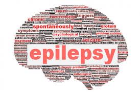
Epilepsy is a central nervous system disorder that causes seizures which affect various mental and physical functions like changes in attention and behavior. This disorder disturbs nerve cell activity in your brain which in turn leads to abnormal behavior, symptoms and sensations involving loss of consciousness.

Symptoms of seizures during epilepsy vary from person to person. Not all the people who have experienced seizures once in a life time are suffering from epilepsy; epilepsy is a group of related disorders; where a patient develops a tendency towards recurrent seizures.


What are the causes of Epilepsy?

In majority of the cases no discernible cause of epilepsy can be traced out. In some cases, it occurs due to some sort of brain damage. Any damage to brain can disturb its normal functioning and lead to seizures. The potential causes of seizures are:

  • In elderly people, epilepsy can occur due stroke, Alzheimer’s disease, head trauma
  • Traumatic brain injury
  • Infections like; Meningitis, Brain Abscess, AIDS
  • Genetic Brain problems
  • Birth defects
  • Problems during birth which deprived the child of oxygen like; twisting or compressing of umbical cord during birth
  • Brain Injuries occurred during or near birth
  • Brain Tumors
  • Abnormal blood vessels of brain
  • Any disease that can damage brain
  • Using certain medicines like; anti-depressants, tramdol etc.
  • Cerebral palsy
  • Drugs and Misuse of alcohol

Which are the potential triggers of Epileptic seizures?

In certain patients, some specific substances or circumstances can trigger seizures. These involve:

  • Stress
  • Insufficient Sleep
  • Over consumption of Alcohol
  • Using illegal drugs, such as; cocaine, ecstasy
  • Opiate based drugs like- heroine, methadone
  • Poor health conditions leading to high fever
  • Flashing and flickering light

What are the symptoms of Epilepsy?

Symptoms of Epilepsy depend upon the kind of seizures a patient is experiencing. The symptoms which a patient exhibits generally depend upon the part of the brain affected and the reason of epilepsy as well. These are:

  • Generalized Seizures: Generalized seizures occur when all the areas of brain (the cortex) are involved. The person experiencing these seizures:
  1. May cry out
  2. Make some kind of noises
  3. Stiffen for several seconds to minutes
  4. Violent shaking of arms and legs
  5. Dilated eyes
  6. The patient appears not to be breathing and generally turns blue
    1. Noisy breathing
    2. Biting tongue
  • Partial or Focal Seizures: Patients with focal seizures may exhibit the following symptoms:
    1. Abnormal muscle contraction
    2. Normally one side of the body gets affected
    3. Forced turning of the head
    4. Staring spells
    5. Abnormal mouth movements
    6. Lip Smacking
    7. Repetitive and Compulsive actions
    8. Chewing without cause
    9. Tingling sensation
    10. Hallucinations
    11. Abdominal pain
    12. Nausea or Sweating
    13. Black out spells
    14. Mood Swings
  • Absence Seizures: These types of seizures occur only for few seconds and involve the following symptoms:
  1. Occur may times a day
  2. Interfere with school or learning
  3. Misinterpreted as lack of attention or misbehavior
  4. Patients may stop walking or stop talking in the mid of the sentence and starts again
  5. Fumbling of hands
  6. Lack of alertness about the surroundings

What are the Auras in Epilepsy?

People suffering from epilepsy often get distinctive warning signs before experiencing seizures, these are known as auras. These may vary from person to person and involve:

  • Observing strange smell or taste
  • Feeling of déjà vu
  • Feeling that world is dreamlike or unreal
  • Feeling of fear or anxiety

What are the treatments available for Epilepsy?

The most of the patients suffering from epilepsy are treated using drug therapy. The treatment usually depends upon the age, overall health, medical history and as well as recurrence and severity of symptoms. The common options available are:

  • If epileptic seizures are occurring due to brain tumor, abnormal blood vessels in brain or bleeding in brain, doctors may recommend surgery.
  • Epileptic seizures can be treated with the help of medications known as anti consultants. But be cautious as you can not switch the quantity or dosage of medications at your own will. Consult your doctor before taking any such decision.
  • Children suffering form epilepsy disorder are put on Ketogenic diet. This type of diet contains high quantity of fats and low rates of carbohydrates. Children put on Ketogenic diets respond well to this treatment therapy and after few years of practice may stop that diet and remain seizure free.
  • Another treatment technique employed by doctors is Vagus Nerve Stimulation technique; where a vagus nerve stimulator is implanted under your chest skin and its wires are connected to the vagus nerve in your neck. The device sends bursts of electric energy to brain via vagus nerve and helps to lessen the seizures.

Contact Braner Pain Clinic for the effective and timely diagnosis and treatment of any of the neurological disorders. Our professional and talented team of physicians is efficient to handle any neurological disability and make you as staunch and sturdy as before.

Osteoporosis And You

What is OSTEOPOROSIS?

Osteoporosis is a bone disease, that lowers the bone mineral density (BMD) in bones, thus leading to bone fracture. This disease weakens the bones so much so that even a minor fall, stress, strain or jerk; breaks or fractures the bone. The bones of hip, spine and wrist are most commonly impaired and harmed by this disease. Other than these, at times, bones of arms and pelvis are also get affected.

Factors causing Osteoporosis:

Various manageable and unmanageable conditions collectively put a person at the risk of getting affected by Osteoporosis.

Unmanageable Conditions include :

  • Age : The older you get, the higher is the risk of getting osteoporosis.
  • Gender : Usually women are at higher risk than men.
  • Body Structure : Women with small and thin body structure have greater risk.
  • Racial Background : White and Asian women are generally affected by this disease.
  • Family Background : People with osteoporosis background have the greater chances of getting the same.
  • Low Estrogen Level : Women suffering from low estrogen level due to missed menstrual cycle or post menopause comes under the risk group.
  • Medicines : Prolonged use of medicines treating arthritis, diabetes, asthma and cancer can cause this disease.

Manageable Conditions include :

  • Eating Disorders : Eating disorders like; anorexia nervosa i.e. eating less food than required or not eating enough food and veggies lead to osteoporosis.
  • Vitamin D and Calcium Defficiency: Eating calcium and vitamin D deficient diet can lead to bone loss.
  • Less Activity : Lack of exercise, low activity and prolonged bed rests can weaken the muscles.
  • Excessive Smoking and Alcohol : Smoking and in taking too much of alcohol can cause weak and broken bones.

Symptoms: Generally, there are no warning signals in the initial stages but once the bones get weakened you may experience :

  • Sudden back pain due to fracture
  • Loss of height
  • Stooped or hunched posture or back
  • Bone fracture due to minor fall or stress

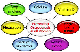
 Prevention : By following the below mentioned easy tips and steps you can control and as well can prevent osteoporosis :

Prevention from Osteoporosis

Prevention from Osteoporosis

  • Eat Calcium Rich Diet : Intake sufficient amount of calcium in your diet, in form of food and nutritional supplements. Foods like milk, plain or fat free yogurt, spinach and white beans can enrich your diet with calcium.
  • Consume sufficient Vitamin D : Vitamin D is produced in our body by exposing our skin to direct sunlight or by consuming Vitamin D rich foods such as ; eggs, milk and salmon. You can also intake vitamin D in the form of nutritional supplements or pills.
  • Opt for Nutritional Diet : Take foods that are rich sources of vitamin K, vitamin C, Zinc and Magnesium as well.
  • Exercise : Regularly practice weight bearing and general exercises mentioned below to strengthen and mobilize your muscles :
    1. Walking or Jogging
    2. Dancing
    3. Running
    4. Ascending and Descending Stairs
    5. Hiking
    6. Playing Tennis and Lifting Weights
    7. Practice Yoga
  • Quit Smoking : Smoking along with impairing your heart and lungs, also damages your bones and as well lead to bone loss by reducing the amount of estrogen in your body.
  • Drink reasonably and sensibly : Have one alcoholic drink per day. Excessive drinking instabilizes your balance and as well affects the calcium absorption capacity of your body.

In order to prevent falls and fractures, you can make your home safer and hurdle free by following the below mentioned instructions : 

  1. Make use of  a cane stick or walker for walking
  2. Use rubber soled shoes and avoid walking in stockings or slippers
  3. Use your grass path ways if your side walks are slippery and wet
  4. Clutter free your room
  5. Always spread skid proof rugs and carpets

Treatment :

 In order to prevent fractures in future due to osteoporosis, along with medications; changes in lifestyle, a calcium, vitamins and minerals enriched diet and regular exercising  is also crucial and important. Before prescribing any drugs or medications to treat and cure osteoporosis, health care practitioners generally consider; the age, gender and severity of condition. To cure and recover you from fracture , a  physical therapist may  :

  1. Use relaxation techniques to relieve muscle tension
  2. Apply hot or cold treatments using warm baths and cold packs to relieve pain
  3. Administer TENS therapy, where nerves are stimulated to relieve pain

Contact Braner Pain Clinics, even if after making all the necessary efforts, you are still suffering from the pain caused due to osteoporosis. We offer holistic treatment options to cure  and treat  your pain and also help you to live your life to the fullest.  

Neurological Evaluation and Treatment

Neurological Evaluation

Neurological Evaluation is a diagnostic procedure, conducted to determine any peripheral neuropathy i.e. neurological impairment in a patient. Physical examination and review of patient’s medical history together make up for neurological assessment. Inferences drawn are based upon the findings of both the tests conducted. Doctors use it both as a screening tool and investigative tool as well.

Neurological Testing and Neurological Disability Clinic in Virginia

Neurological Testing and Neurological Disability Clinic in Virginia

Steps Involved

 Neurological Evaluation is carried out in several steps. These steps are not necessarily followed in the same pattern i.e. physicians can perform them randomly. Below stated steps are followed  while administering a neurological evaluation :

  • Higher Functions or Mental Status Examination
  • Cranial Nerves Examination
  • Sensory System Examination
  • Motor  System Examination
  • Deep Tendon Reflexes or Reflexes’ Examination
  • Cerebellum
  • Meninges
  • System Survey

Tests Administered

 Various tests including the following are conducted to detect the root cause of any neurological disorder and on the basis of the results drawn out, suitable treatment options are forwarded :

  • Review of Medical History : While administering this test physicians ask questions related to the onset of symptoms and associated discomfort, age gender occupation of the patient, hands he use to perform functions( right or left hand), his past medical and drug history along with his family and social history. Moreover physical exam is performed to measure his reflexes and sensations.
  • Neurological Evaluation : Tests are conducted to assess patient’s muscle strength and functioning of his autonomic nerves along with his sensory experience.
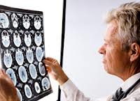
  • Diagnostic Testing : Depending upon the condition and need and necessities of the patients; nerve biopsy, spinal tap or magnetic resonance imaging (MRI) is recommended and performed by the neurologist.
  • Electro diagnostic Tests :  To measure the electric activity of muscles and nerves, electro diagnostic tests are administered. These tests further help to determine nerve damage, its cause and extent.
  • Blood Tests : To detect the toxic elements and  vitamin deficiencies and moreover to trace an impaired immune response doctors advice laboratory tests for the following :
  • Vitamin B12 and folate levels
  • Kidney, Liver and Thyroid functioning
  • Oral Glucose tolerance Test
  • Lyme Disease
  •  Hepatitis B and C

Contact Braner Pain Clinics

If you are facing any of the neurological symptoms or discomforts, Consult Braner Pain Clinics for their comprehensive and sophisticated evaluation, diagnosis and treatment.  We specialize in the area of neurological evaluation and chronic pain management.

Myasthenia Gravis

Clinical Features of Myasthenia Gravis:

Myasthenia GravisMyasthenia gravis is a disorder of neuromuscular transmission characterised by:

  • Weakness and fatiguing of some or all muscle groups
  • Weakness worsening on sustained or repeated exertion, or towards the end of the day, relieved by rest

This condition is a consequence of an autoimmune destruction of the NICOTIN1C POSTSYNAPTIC RECEPTORS FOR ACETYLCHOLINE.

Myasthenia gravis is rare, with a prevalence of 5 per 100 000. The increased incidence of autoimmune disorders in patients and first degree relatives and the association of the disease with certain histocompatibility antigens (HLA) – B7, B8 and DR2 – suggests an IMMUNOLOGICAL BASIS.

Antibodies bind to the receptor sites resulting in their destruction (complement mediated). These antibodies are referred to as ACETYLCHOLINE RECEPTOR ANT1BODIES (AChR antibodies) and are demonstrated by radioimmunoassay in the serum of 90% of patients.

Muscle biopsy may show abnormalities:

  • Lymphocytic infiltration associated with small necrotic foci of muscle fibre damage.
  • Muscle fibre atrophy (type I and II or type III alone).
  • Diffuse muscle necrosis with inflammatory infiltration (when associated with thymoma)

Motor point biopsy may show abnormal motor endplates. Supravital  methylene blue staining reveals abnormally long and irregular terminal nerve branching. Light and electron microscopy show destruction of ACh receptors with simplification of the secondary folds of the postsynaptic surface.

Clinical Features

Up to 90% of patients present in early adult life (<40 years of age). Female: male ratio 2:1. The disorder may be selective, involving specific groups of muscles.

Several clinical subdivisions are recognised:

  1. Class 1 – ocular muscles only – 20%
  2. Class 2 – Mild generalised weakness
  3. Class 3 – Moderate generalised and mild to moderate ocular-bulbar weakness
  4. Class 4 – Severe generalised and ocular-bulbar weakness
  5. Class 5 – Myasthenic crises

Approximately 40% of class I will eventually become widespread. The rest remain purely ocular throughout the illness. Respiratory muscle involvement accompanies severe illness.

Clinical Features

Cranial nerve signs and symptoms

  • Ocular involvement produces ptosis and muscle paresis.
  • Weakness of jaw muscles allows the mouth to hang open.
  • Weakness of facial muscles results in expressionless appearance.

Braner Pain Clinics has a talented and friendly staff. We will do everything in our power to make sure your visit is a satisfying experience. If there is anything else you may need from us, just ask! We are here to serve you.

How to Recover from Spinal Cord Disease?

Clinical Features and Management of Spinal Cord Disease:

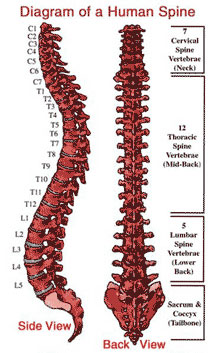
spine-diagramIn developing countries, spinal TB is mostly a disease of childhood or adolescence. It usually affects the middle aged and is particularly prevalent in immigrant populations and in the immunocompromised. The incidence is now increasing, probably due to the development of antibiotic resistance.

The lower thoracic spine is commonly involved and the disease initially affects the intravertebral disc and spreads to adjacent vertebral bodies.

Clinical Features:

  • The classic systemic features of weight loss, night fever and cachexia are often absent.
  • Pain occurs over the affected area and is made worse by weight bearing.
  • Symptoms and signs of cord compression occur in approximately 20% of cases.
  • The onset may be gradual as pus, caseous material or granulation tissue accumulate, or sudden as vertebral bodies collapse and a kyphosis develops.

Straight X-rays are characteristics

  • Anterior superior or inferior angle of the vertebral body is initially involved.
  • Infective process spreads throughout the vertebral body and may involve the pedicles or facet joints.
  • The disc space collapses as the vertebral plate is destroyed.

MRI with gadolinium shows an epidural mass with paraspinal soft tissue swelling.

Management:

Every effort is made to establish the diagnosis. A needle biopsy is often sufficient, but occasionally an exploratory operation (costotransversectomy) is required. Long-term antituberculous therapy is commenced.

If signs of cord compression develop, decompression is necessary.

  • A Posterior Decompression, removing the remaining unaffected bone, is likely to cause instability an anterior or posterolateral approach is therefore required.
  • Posterolateral Approach (costotransversectomy): One or more ribs are resected medially, along with the transverse processes.
  • Anterior Transthoracic Decompression with strut graft fusion is something performed. This permits clearance of pus and caseous debris without retracting the spinal cord.

Braner Clinics is a professional corporation, and was established for the practice of chronic pain management in 1990. For best treatment of Spinal Cord Disease call now for Quick Appointment: (703) 573-1282

What is Cerebral Aneurysms?

Investigation and Treatment of Cerebral Aneurysms:

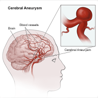
At autopsy intracranial aneurysms are found in approximately 2% of the population. Aneurysm rupture occurs in 6 -8 per 100 000 per year.

Inheritance:

Investigations reveal aneurysms in 10% of relatives with two or more affected 1st degree family members. The genetic basis remains unknown. Procollagen III deficiency may play a role in some patients.

Cerebral Aneurysm

Morphology

  • Intracranial aneurysms are usually saccular, occurring at vessel bifurcations. Size varies from a few millimetres to several centimetres. Those over 2.5 cm are termed ‘giant’ aneurysms.
  • Fusiform dilatation and ectasia of the carotid and the basilar artery may follow atherosclerotic damage. These aneurysms seldom rupture. Mycotic aneurysms, secondary to vessel wall infection, arise from haematogenous spread, e.g. subacute bacterial endocarditis.
  • Aneurysm rupture usually occurs at the fundus of the aneurysm and the risk is related to size. Smoking, hypertension and alcohol excess also play a part. In some patients, rupture occurs during exertion, straining or coitus, but in most there is no associated relationship.
  • Multiple aneurysms: In approximately 30% of patients with aneurysmal SAH, more than one aneurysm is demonstrated on angiography.

Pathogenesis

  • The cause of aneurysm formation may be multifactorial with acquired factors combining with an underlying genetic susceptibility.
  • Aneurysms were once thought to be congenital due to the finding of developmental defects in the tunica media.
  • These defects occur at the apex of vessel bifurcation as do aneurysms, but they are also found in many extracranial vessels as well as intracranial vessels; saccular aneurysms in contrast are seldom found outwith the skull.
  • Tunica media defects are often evident in children, yet aneurysms are rare in this age group.
  • It now appears that defects of the internal elastic lamina are more important in aneurysm formation and these are probably related to arteriosclerotic damage.
  • Aneurysms often form at sites of haemodynamic stress where for example, a congenitally hypoplastic vessel leads to excessive flow in an adjacent artery. It is not known whether they form rapidly over the space of a few minutes, or more slowly over days, weeks, or months.
  • Hypertension may play a role; more than half the patients with ruptured aneurysm have pre-existing evidence of raised blood pressure.

Clinical Presentation

Of those patients with intracranial aneurysms presenting acutely, most have had a subarachnoid haemorrhage. A few present with symptoms or signs due to compression of adjacent structures. Others present with an aneurysm found incidentally.

  1. Rupture
  2. Compression from aneurysm sac
  3. Incidental finding

Investigation

All patients deteriorating suddenly require a CT scan. This helps in establishing the diagnosis of rebleeding and excludes a remediable cause of the deterioration, e.g. acute hydrocephalus.

Treatment

Aneurysm treatment requires a team approach involving interventional radiologists and neursurgeons. Treatment selection must take a variety of factors into account including the nature and location of the aneurysm, the relative difficulties of the endovascular or operative approach and the patients age and clinical condition.

Braner Pain Clinics has a talented and friendly staff. We will do everything in our power to make sure your visit is a satisfying experience. If there is anything else you may need from us, just ask! We are here to serve you.

http://www.branerpainclinic.com

Are You At Risk For Osteoporosis?

Clinical Features and Treatment of Osteoporosis:

OsteoporosisOsteoporosis is characterised by abnormal reduction in bone mass per unit volume, due to poor formation of organic protein matrix. The total bone mass is diminished but the bone present is qualitatively normal. Osteoporosis results because the rate of bone resorption is higher than that of bone formation. There occurs marked reduction in the mechanical strength of the bone and makes the bone vulnerable for fractures following a trivial trauma.

Osteoporosis is a disease of the elderly. It is seen more commonly in females.

Types of Osteoporosis

Osteoporosis can be categorised into two types.

  • Primary
  • Secondary

Primary osteoporosis

  • Type I-postmenopausal osteoporosis: In females, the hormonal changes at menopause cause rapid loss of trabecular bone and result into generalised osteoporosis. These patients became susceptible to fractures of the vertebrae, neck of femur and distal radius.
  • Type II-senile osteoporosis: This affects men and women over 60 years of age.

Secondary osteoporosis

A variety of conditions/factors can cause osteoporosis secondarily. These can be broadly classified as:

1.       Endocrinal

  • Cushing’s syndrome
  • Hyperthyroidism

2.       Nutritional

  • Malnutrition

3.       Drug induced

  • Prolonged cortisone use

4.       Immobilisation

  • In fractures
  • Bed-ridden patients

5.       Miscellaneous

  • Cancer disease
  • Multiple myeloma

Clinical Features

  • The patient presents with backache and pain in the bones.
  • The patient gradually develops progressive kyphosis of the dorsal spine.
  • More commonly, the patients present with a fracture of neck of femur or the distal radius.
  • On examination, there is diffuse tenderness over the spine and other bones generally.
  • However, the tenderness may be localised over the spine if there is a pathological compression fracture of the vertebrae.
  • There is some decrease in the height due to kyphosis.

Treatment

The mainstay of the treatment is prevention of osteoporosis and measures to increase the bone density, thereby reducing the risk of fractures.

1.     Good nutritious diet: This is important because malnutrition and dietary deficiencies are associated with osteoporosis.

2.  Exercise: A good level of physical activity acts as an important stimulus to the skeleton; and disuse, immobilisation or denervation paralysis results in bone loss. Physical exercise for 45 minutes at least four times a week helps in increasing the bone mass.

3.  Calcium: It has been seen that intake of additional calcium may reduce the bone loss, particularly in postmenopausal women. The recommended dose is 1000 to 1500 mg/day.

4.    Vitamin D: Deficiency of vitamin D is commonly seen in the elderly and leads to secondary hyperparathyroidism with resultant bone loss. It helps in the absorption of calcium from the gut.

5.     Hormone replacement therapy (HRT): In postmenopausal osteoporosis, HRT reduces the incidence of vertebral and hip fractures.

6.  Selective oestrogen receptor modulation (SERM) is a newer class of agent which has effects similar to those of low doses of oestrogen. Raloxifene is a commercially available SERM which is generally well tolerated with a decreased incidence of breast cancer; unlike HRT.

7.    Calcitonin: Calcitonin is a peptide hormone secreted by the C-cells of thyroid. It directly inhibits bone resorption. It is known to reduce the incidence of fractures by improving the bone mass. It is administered by injections, but nowadays intranasal preparations are also available.

Your bones won’t tell you if they are weak. In fact you may have osteoporosis and never know it. For many people, breaking a bone is their first clue. Call now for Quick Appointment: 1 (877) 573-1282
http://www.branerpainclinic.com

Results and complications of Facial Pain

Investigation and Treament of Facial Pain:

Trigeminal-NeuralgiaPain in the face may result from many different disorders and often presents as a diagnostic problem to the neurologist or neurosurgeon.

Quality of pain

  • Trigeminal neuralgia – sharp, stabbing, shooting, paroxysmal
  • Atypical facial pain – dull, persisting
  • Postherpetic neuralgia – dull, burning, persisting, occasional paroxysm
  • Dental – dull
  • Sinusitis – sharp, boring, worse in the morning
  • Ocular – dull, throbbing
  • Costen’s syndrome – severe aching, aggravated by chewing
  • Cluster headache – sharp, intermittent

Trigeminal Neuralgia

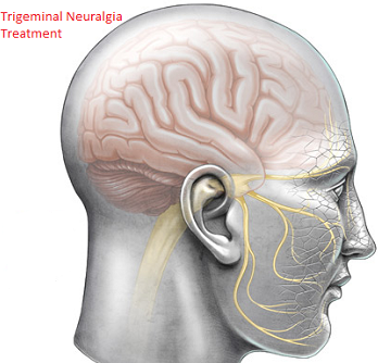
Trigeminal neuralgia is characterised by paroxysmal attacks of severe, short, sharp, stabbing pain affecting one or more divisions of the trigeminal nerve. The pain involves the second or third divisions more often than the first; it rarely occurs bilaterally and never simultaneously on each side, occasionally more than one division is involved. Paroxysmal attacks last for several days or weeks; they are often superimposed on a more constant ache. When the attacks settle, the patient may remain pain free for many months.

Chewing, speaking, washing the face, tooth-brushing, cold winds, or touching a specific ‘trigger spot’, e.g. upper lip or gum, may all precipitate an attack of pain.

Trigeminal neuralgia more commonly affects females and patients over 50 years of age.

Aetiology

Trigeminal pain may be symptomatic of disorders which affect the nerve root or its entry zone

Root or root entry zone compression

  • Arterial Vessels often abut and sometimes clearly indent the trigeminal nerve root at the entry-zone into the pons, causing ephaptic transmission (short circuiting).
  • Tumours of the cerebellopontine angle lying against the V nerve roots, e.g. meningioma, epidermoid cyst, frequently present with trigeminal pain.

Demyelination – such a lesion in the pons should be considered in a ‘young’ person with trigeminal neuralgia. Trigger spots are rare. Remission occurs infrequently and the response to drug treatment is poor.

In some patients the cause remains unexplained, as do the long periods remission.

Investigation

MR scan to exclude a cerebello-pontine angle lesion or demyelination.

Results and complications

Pain relief- no comparative trials have been done so accurate comparison of the wide variety of techniques used for trigeminal neuralgia is difficult. Microvascular decompression seems to be more likely to provide pain control with fewer relapses. Overall 80-85% of patients remain pain free for a 5-year period. Results of peripheral nerve avulsion are less satisfactory with pain recurring in 5000 within 2 years.

  • Dysaesthesia\Anaesthesia dolorosa – This troublesome sensory disturbance follows any destructive technique to nerve or root in 5-30% of patients. Microvascular decompression avoids this.
  • Corneal anaesthesia – This occurs when root section or thermocoagulation involves the first division and keratitis may result.
  • Mortality – Microvascular decompression and open root section carry a very low mortality (< 1%), but this must not be ignored when comparing results with safer methods.

Treatment selection: This depends on discussion of the differing risks with the patient. In younger patients the absences of sensory complications make microvascular decompression the procedure of first choice. Frail and elderly patients may tolerate glycerol injection, balloon compression and thermocoagulation more easily than other procedures.

Braner Clinics is a professional corporation, and was established for the practice of chronic pain management in 1990. Appointments are on a first come first serve basis. If you are going to miss your appointment please call at least 48 hours in advance, as there may be a waiting list. You may be asked for a credit card deposit for $25 towards your appointment. This deposit will be deducted from your first visit. If you miss your appointment this deposit will not be refunded to you.

Management of Spinal Cord Injury

Information about Spinal Trauma (Injury):

Spinal TraumaSpinal Trauma is due to harm to the vertebrae that results in a bruise (also known as a contusion), a limited split, or a complete split. Because the vertebra is the main service provider of alerts throughout the body, harm to the vertebrae. At impact, spinal cord damage may or may not accompany the bony or ligament damage.

After impact, stability at the level of injury plays a crucial part in further management. Injudicious movement of a patient with an unstable lesion may precipitate spinal cord injury or aggravate any pre-existing damage.

Management

Management depends on the site and stability of the lesion, but basic principles apply.

  • An unstable lesion risks further damage to the spinal cord and roots and requires either operative  fixation or immobilisation, e.g. skull traction, Halo or plaster jacket.
  • There is no evidence that ‘decompressing’ the cord lesion (either anteriorly or posteriorly) improves the neurological outcome.
  • If patients with normal cord function or with an incomplete cord lesion (i.e. with some residual function) progressively deteriorates, then operative decompression is required.

Many additional techniques (e.g. steroids, cord cooling, and hyperbaric oxygen) have been employed with the aim of improving neurological outcome.

Management of the Paraplegic Patient

After spinal cord injury, transfer to a spinal injury centre with medical and nursing staff skilled in the management of the paraplegic patient provides optimal daily care and rehabilitation.

Important Features Include:

  1. Skin Care
  2. Urinary Tract
  3. Limb

Outcome Following Spinal Cord or Root Injury

  • Patients with high cervical vertebral cord lesions seldom survive without immediate ventilatory support.
  • Patients who survive a lesion above C7 usually remain dependent on others for daily care.
  • Patients with thoraco-lumbar injuries usually regain full independence.
  • A mixed cord and lumbar root lesion may occur at this level. Fortunately roots are more resistant to injury ‘root escape’ and the outlook is more favourable.

Braner Clinics is a professional corporation, and was established for the practice of chronic pain management in 1990. Pain Management is the specialty of PAIN MEDICINE for the assessment, diagnosis and care of severe, chronic persistent pain conditions and human suffering that persists beyond the reasonable, expected healing time after injuries or illnesses, usually after 3 months. Call now at: (877) 573-1282 or Visit our Website: http://www.branerpainclinic.com