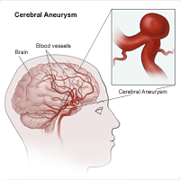
Investigation and Treatment of Cerebral Aneurysms:
At autopsy intracranial aneurysms are found in approximately 2% of the population. Aneurysm rupture occurs in 6 -8 per 100 000 per year.
Inheritance:
Investigations reveal aneurysms in 10% of relatives with two or more affected 1st degree family members. The genetic basis remains unknown. Procollagen III deficiency may play a role in some patients.
Morphology
- Intracranial aneurysms are usually saccular, occurring at vessel bifurcations. Size varies from a few millimetres to several centimetres. Those over 2.5 cm are termed ‘giant’ aneurysms.
- Fusiform dilatation and ectasia of the carotid and the basilar artery may follow atherosclerotic damage. These aneurysms seldom rupture. Mycotic aneurysms, secondary to vessel wall infection, arise from haematogenous spread, e.g. subacute bacterial endocarditis.
- Aneurysm rupture usually occurs at the fundus of the aneurysm and the risk is related to size. Smoking, hypertension and alcohol excess also play a part. In some patients, rupture occurs during exertion, straining or coitus, but in most there is no associated relationship.
- Multiple aneurysms: In approximately 30% of patients with aneurysmal SAH, more than one aneurysm is demonstrated on angiography.
Pathogenesis
- The cause of aneurysm formation may be multifactorial with acquired factors combining with an underlying genetic susceptibility.
- Aneurysms were once thought to be congenital due to the finding of developmental defects in the tunica media.
- These defects occur at the apex of vessel bifurcation as do aneurysms, but they are also found in many extracranial vessels as well as intracranial vessels; saccular aneurysms in contrast are seldom found outwith the skull.
- Tunica media defects are often evident in children, yet aneurysms are rare in this age group.
- It now appears that defects of the internal elastic lamina are more important in aneurysm formation and these are probably related to arteriosclerotic damage.
- Aneurysms often form at sites of haemodynamic stress where for example, a congenitally hypoplastic vessel leads to excessive flow in an adjacent artery. It is not known whether they form rapidly over the space of a few minutes, or more slowly over days, weeks, or months.
- Hypertension may play a role; more than half the patients with ruptured aneurysm have pre-existing evidence of raised blood pressure.
Clinical Presentation
Of those patients with intracranial aneurysms presenting acutely, most have had a subarachnoid haemorrhage. A few present with symptoms or signs due to compression of adjacent structures. Others present with an aneurysm found incidentally.
- Rupture
- Compression from aneurysm sac
- Incidental finding
Investigation
All patients deteriorating suddenly require a CT scan. This helps in establishing the diagnosis of rebleeding and excludes a remediable cause of the deterioration, e.g. acute hydrocephalus.
Treatment
Aneurysm treatment requires a team approach involving interventional radiologists and neursurgeons. Treatment selection must take a variety of factors into account including the nature and location of the aneurysm, the relative difficulties of the endovascular or operative approach and the patients age and clinical condition.
Braner Pain Clinics has a talented and friendly staff. We will do everything in our power to make sure your visit is a satisfying experience. If there is anything else you may need from us, just ask! We are here to serve you.
